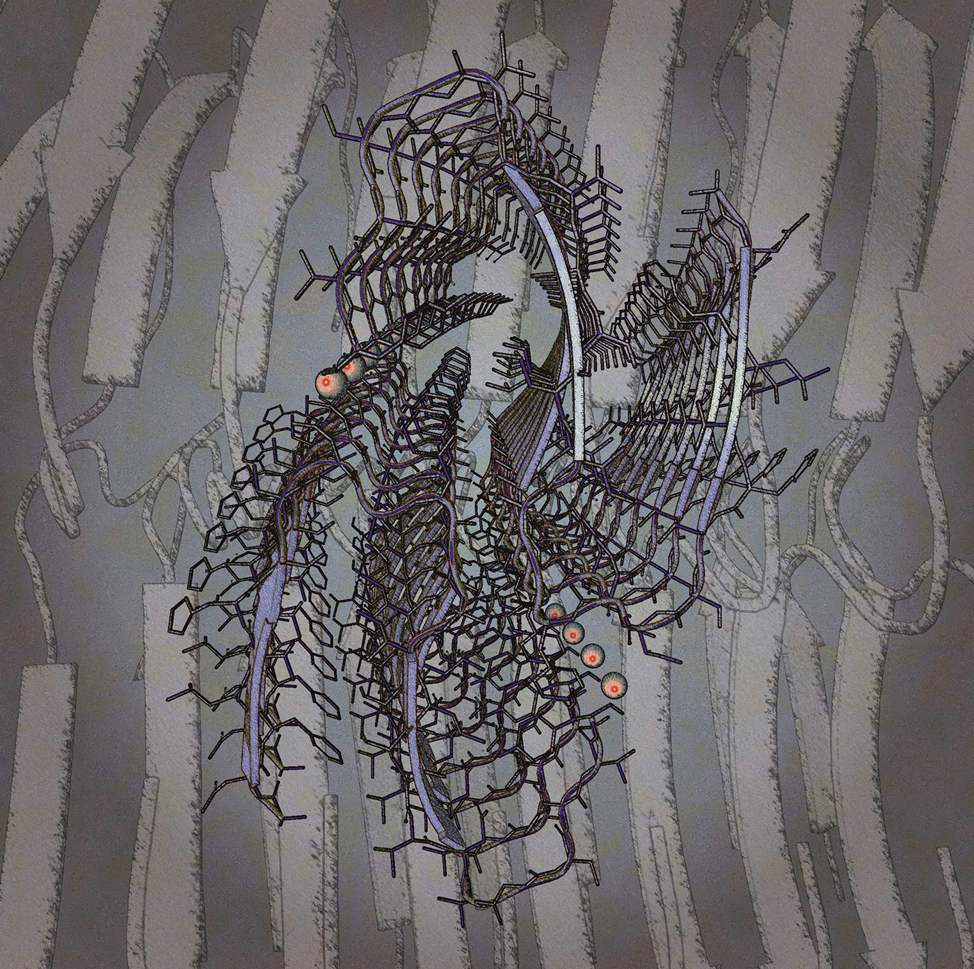


Amyloids are aggregates of proteins characterised by a fibrillar morphology of 7–13 nm in diameter, a β-sheet secondary structure (known as cross-β) and ability to be stained by particular dyes, such as Congo red. In the human body, amyloids have been linked to the development of various diseases. Pathogenic amyloids form when previously healthy proteins lose their normal structure and physiological functions (misfolding) and form fibrous deposits in plaques around cells which can disrupt the healthy function of tissues and organs. Such amyloids have been associated with (but not necessarily as the cause of) more than 50 human diseases generally known as amyloidosis, and typically related with a cognitive deficiency in the central nervous system. Amyloids have been known to arise from many different proteins. These polypeptide chains generally form β-sheet structures that aggregate into long fibers; however, identical polypeptides can fold into multiple distinct amyloid conformations. Here you can see the structure of an amyloid formed by the islet amyloid polypeptide (IAPP). The formation of these amyloid fibrils in the pancreatic cells has been associated with beta-cell loss and have been implicated in type 2 diabetes (T2D). The structure was solved by CryoEM using an helical reconstruction protocol (PDB code: 6Y1A)
#molecularart ... #immolecular ... #amyloid ... #diabetes ... #pancreas ... #islet ... #polypeptide ... #cryoem ... #helicalreconstruction
Protein rendered with @proteinimaging and composed by @corelphotopaint
#molecularart ... #immolecular ... #amyloid ... #diabetes ... #pancreas ... #islet ... #polypeptide ... #cryoem ... #helicalreconstruction
Protein rendered with @proteinimaging and composed by @corelphotopaint

